| ACQ4 Neurophysiology Acquisition and Analysis System |
| Electrophysiology - Photostimulation - Fluorescence Imaging |
Screenshots
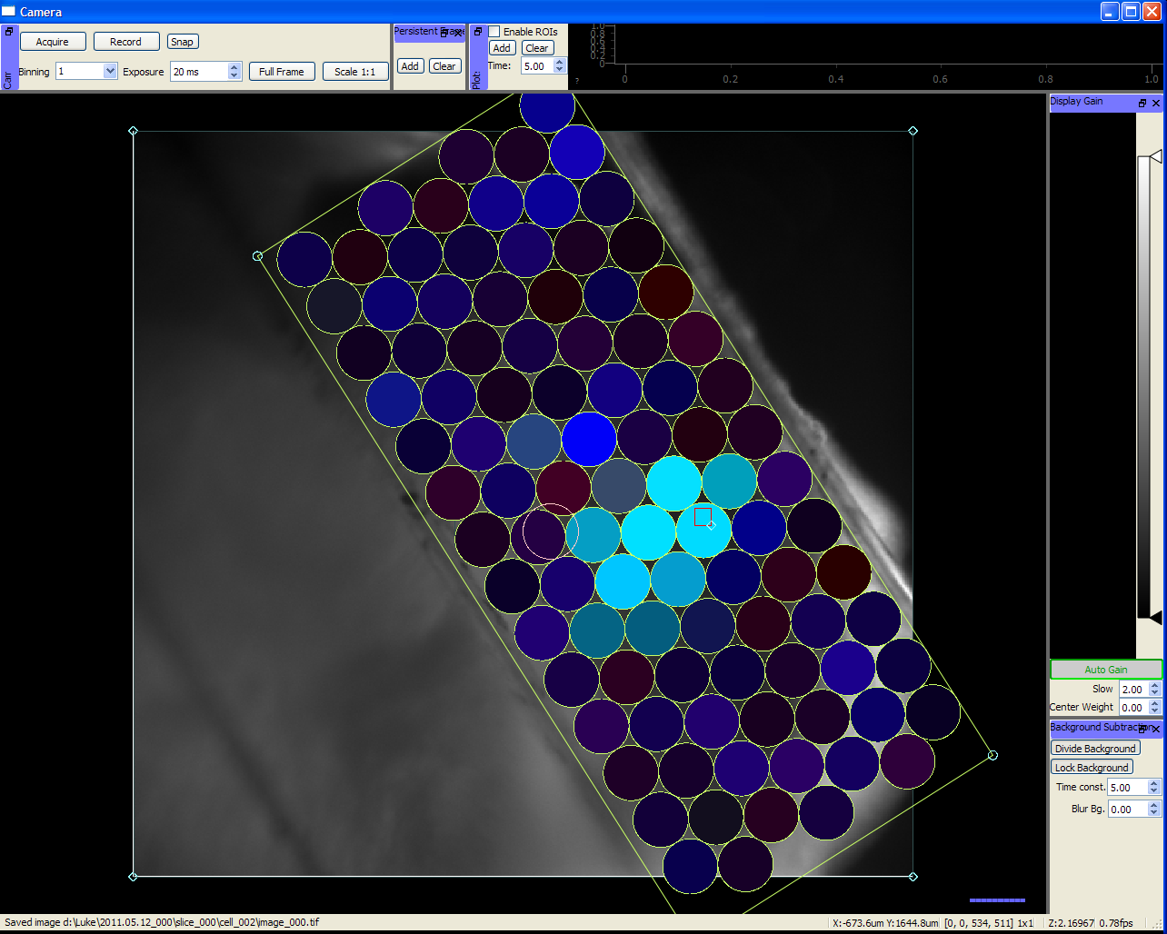
Camera Module - Showing live video feed and online analysis of a photostimulation mapping experiment. The locations to be stimulated are selected by dragging and resizing a grid over the video feed. Results are displayed as each spot is stimulated.
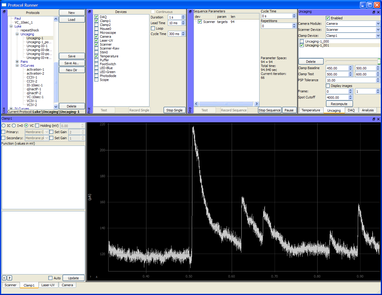
Protocol Runner Module - Showing a photostimulation protocol. Displayed at center is the patch clamp recording for a single stimulation. This protocol synchronizes the patch clamp, camera, laser, scan mirrors and other devices. Each device has its own graphical interface used for configuring the device's behavior during the protocol.
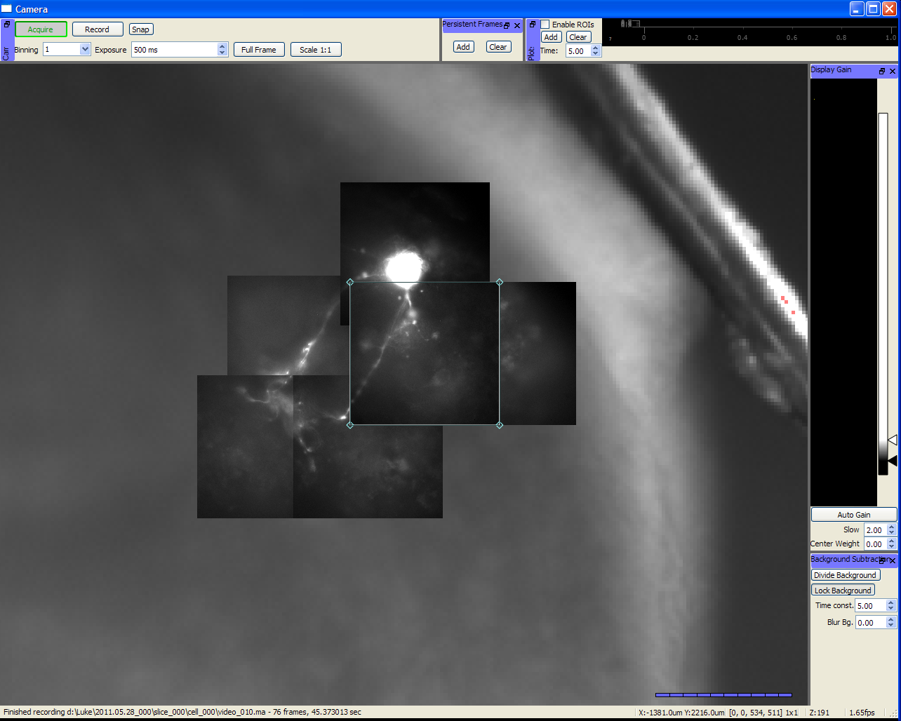
Camera Module - Showing live video feed and online construction of an image mosaic. The video feed moves around the scene as the microscope is moved, and frames are added to the background using the "Add" button above.
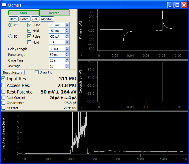
Patch Module - Used to monitor patch progress and long-term cell health. Repeatedly delivers current or voltage pulses to the patch electrode and computes input resistance, access resistance, capacitance, etc.

I/V Curve analysis
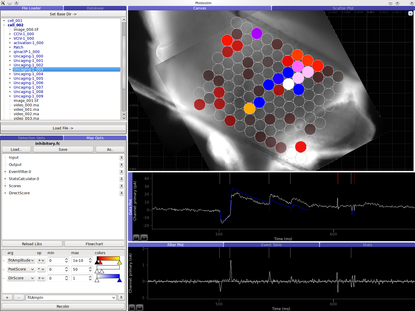
Offline analysis of photostimulation mapping experiment. Shows 1) mosaic of images and spots colored by detection critera, 2) raw waveform displaying detected and normalized presynaptic events, 3) filtered data used for event detection.
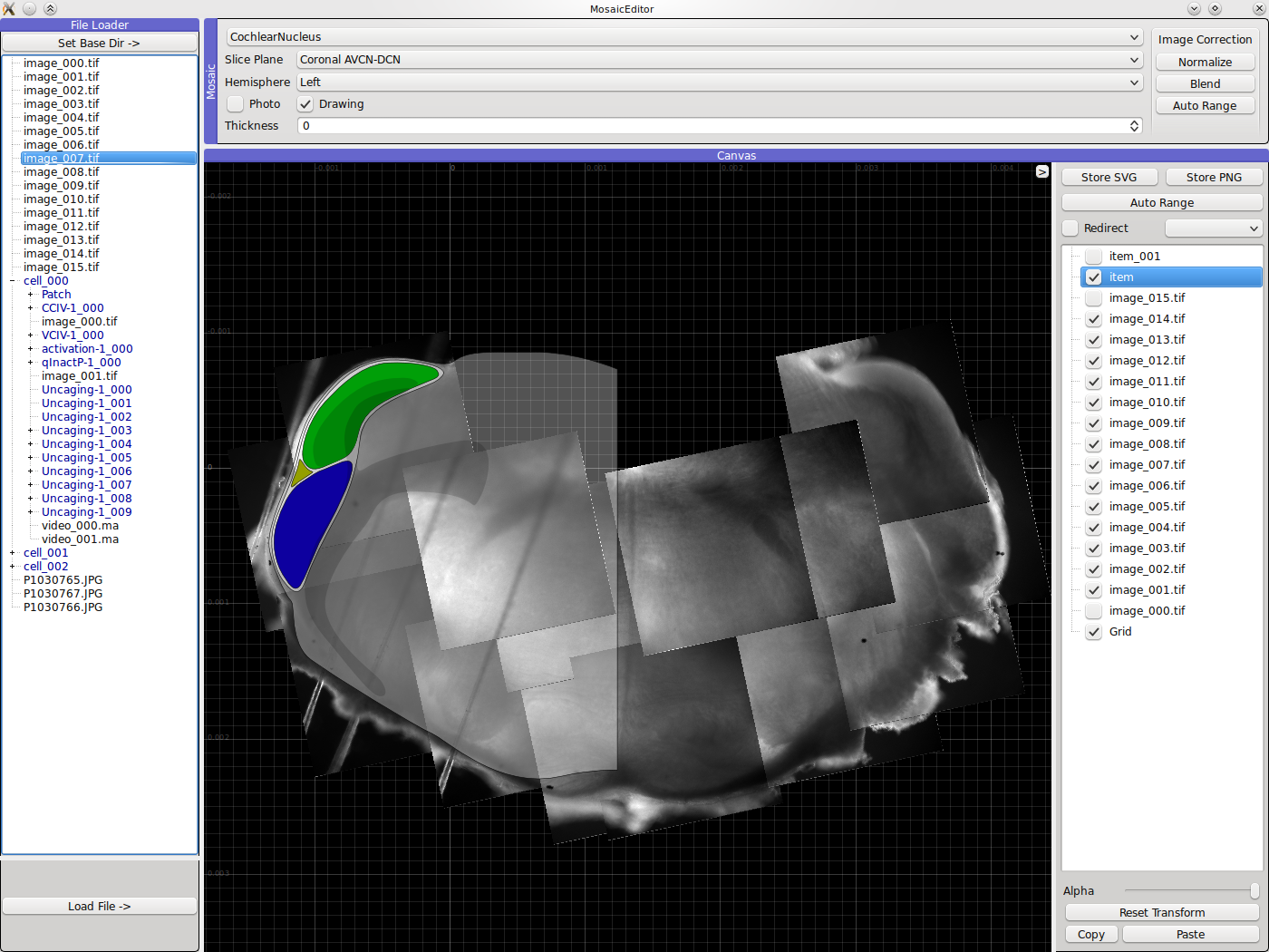
Automatic mosaic reconstruction of slice images taken during an experiment, with anatomical reference overlaid.